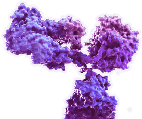
Mouse Anti-ABT1 Antibody (CBMOAB-00193HCB)
To download a Certificate of Analysis, please enter a lot number in the search box below. Note: Certificate of Analysis not available for kit components.
Lot Number
- Product List
- Specifications
- Application Information
- Target
- Relate Reference Data
| Sub Cat | Clonality | Species Reactivity | Application | Clone | Conjugate | Size | |
| CBMOAB-00193HCB | Monoclonal | C. elegans (Caenorhabditis elegans), Cattle (Bos taurus), Chimpanzee (Pan troglodytes), Ferret (Mustela Putorius Furo), Frog (Xenopus laevis), Marmoset, O. mykiss (Oncorhynchus mykiss), Rhesus (Macaca mulatta), Zebrafish (Danio rerio) | WB, ELISA | MO00193HB | 100 µg | ||
| CBMOAB-34900FYA | Monoclonal | Rhesus (Macaca mulatta) | WB, ELISA | MO34900FYA | 100 µg | ||
| CBMOAB-64534FYA | Monoclonal | Zebrafish (Danio rerio) | WB, ELISA | MO64534FYA | 100 µg | ||
| MO-AB-15567W | Monoclonal | Chimpanzee (Pan troglodytes) | WB, ELISA | MO15567W | 100 µg | ||
| MO-AB-34323W | Monoclonal | Ferret (Mustela Putorius Furo) | WB, ELISA | MO34323W | 100 µg | ||
| MO-AB-50275W | Monoclonal | Marmoset | WB, ELISA | MO50275W | 100 µg | ||
| MO-AB-06823R | Monoclonal | Cattle (Bos taurus) | WB, ELISA | MO06823R | 100 µg | ||
| MO-AB-01153H | Monoclonal | Frog (Xenopus laevis) | WB, ELISA | MO01153C | 100 µg | ||
| MO-AB-10596Y | Monoclonal | O. mykiss (Oncorhynchus mykiss) | WB, ELISA | MO10596Y | 100 µg |
Specifications
| Host species | Mouse (Mus musculus) |
| Species Reactivity | C. elegans (Caenorhabditis elegans), Cattle (Bos taurus), Chimpanzee (Pan troglodytes), Ferret (Mustela Putorius Furo), Frog (Xenopus laevis), Marmoset, O. mykiss (Oncorhynchus mykiss), Rhesus (Macaca mulatta), Zebrafish (Danio rerio) |
| Clone | MO00193HB |
| Specificity | This antibody binds to C. elegans ABT1. |
| Format | Liquid or Lyophilized |
| Storage | Store at 4°C: short-term (1-2weeks) Store at -20°C: long-term and future use |
| Purity | > 90% was determined by SDS-PAGE |
| Purification | Purified with Protein A or G affinity chromatography |
Application Information
| Application | WB, ELISA |
| Application Notes | ELISA: 1:1000-1:3000 Other applications are to be developed. The optimal dilution should be determined by the end user. |
Target
| Introduction | Basal transcription of genes by RNA polymerase II requires the interaction of TATA-binding protein (TBP) with the core region of class II promoters. Studies in mouse suggest that the protein encoded by this gene likely activates basal transcription from class II promoters by interaction with TBP and the class II promoter DNA. |
| Product Overview | Mouse Anti-C. elegans ABT1 (clone MO00193HB) Antibody (CBMOAB-00193HCB) is a mouse antibody against ABT1. It can be used for ABT1 detection in Western Blot, Enzyme-Linked Immunosorbent Assay. |
| Alternative Names | Protein ABT-1, isoform a; abt-1 |
| Gene ID | 182849 |
| UniProt ID | S6EZS3 |
| Protein Refseq | The length of the protein is 1558 amino acids long. The sequence is show below: MSSVLSSFSSARRSIRNIWQKDLNYFRSNHPFFILIFIVLSLAFYNVWWHNSPTRDQSGDASKFHLFDNFRLSKQKFVKMIVCIPETVVDYRFSTVIDAIHLTHLDYPHLEFTKKLEKAKNKLEDGEVDFLVLFKTLIETGKKIEYKIISEQGVFDKHCIQCDVHRKKTWGNAVEAQVVVNRFLYWLVKSRGFNKRSPHWQLSNVYVKHSGNDPMFLKRQSDLFLLWILLLPICSRLIDLWNNDRSSGYYNMFLSLHARRFHYFIAKFVFWYGIALIISLIVFIVNVLVVPAVFCSTYLQFVVCYAACIISFAIFLATTFPGSPLFAKIVGFLSMVAGEFGFGTVYHGIGFFQLCVRHFGKSEFDGPWIDAVCTLCMTGNCVLLITVSIYFDTYFCLFTMEPLAFYFPLEKDYWMPEARSPQLDTFFVKQIILLKPGRSEITVKKAHDEILPAKTSTGSKYIPPTPIVKGVPLVVLYGICKRIENHWRVKQVSLTVHLGEVVTLYGHHGCGSGEILSIIAGQMKPEYGEVMMEQSQRPLMISKTTDVPNVNYLNVIQYLRLISKMRGVSASTSHIDEMLEELDLIKVKNRSLDLLSTTQKERLRIAAVFVGQPDLVLIDFPTMECLPEWKFMVFRFIEKRKEKRSIIVSSYDPEETEAISDKVVLMSEGYVVLNGSCEAFKHSINSVFEIRIWPNNRFTDEQIKGMMKTLTLGDNQMKNDARFFETPNGKIRVTLPILYRRSIPLILRELDAVHEKFRIEFYELGKPNIHDIYANACYEHQQLDPLSTFDQLRDHYTKQQPLNKKTYFFKNMIHIFKDGRFLIETAAVVGLFLILTVIILLSYHSVESDSHQTREISLTSYESPITIYCDECEKEVIEGVTFEKKSRVLQLENNQAVLYWKDTAKADKELITVSRGDALFTMIVQNFAVSQMISKITGKHPQIKLFLESENVRTATEISGFSTVFNDISSSKAAMILTESYSLMYITAVFQMFVYTFSVVLPLRLVSTNMGPQSMILPWPRYVYFGFIYVFQLVVFLVIAIILGFAVLSMGFFATNTTSCYLRFLSAWFLSYASTLPLIYFLVFNIQNTKSVIPIILAISSLSVTFPNLVASFSAENQVSLQSLIQFVSWSCMLNPPSSLQVLAAFLNAGESLEKNSGTITSLILFCGAQFWIIVIFVLVIFQPGFSYIQQYWFKSSTVLYSSFERGTTLKPGARIKNSYIFVTNPLGTMEDAQDKGASGNIPNRPAAGTEPVQKPTSDYIAGSSMSIDDMSYITNYEENLVEKMATMTSWNDKQEVPVDNSATFLMGDQTKLKSALLTTIAKINKNEHIAFVPIVENISTIYTPFELLENAAYCNGFEITDDHISYLLDAFDLKEHANTQIKLLPCIDRRILMLLAKLVIKPNIVIIDQLEMFLPHSKMMTVWSFCARLRKNGLGIIYTSHHSGFAEHTATCCAHMYKGQFVRNLLPTSIKAKCTVLEVVPKTASDPKVLLHMLQKEMEESVKLPSTSRNITLAMYKDGVETIEKILELLRMLSPQIKRYSLRSGNCDEFLASAFGK. |
Relate Reference Data

IF
Figure 1 Subcellular localization of mABT1. COS7 cells were transfected with pEGFP-mABT1; after 24 h, cells expressing EGFP proteins were monitored by fluorescence microscopy under phase contrast (A) and dark field (B).


WB
Figure 2 mABT1 interacts with TBP. Coimmunoprecipitation of mABT1 and TBP. COS7 cells were transfected with pcDNA3-myc-mABT1, and lysate was prepared. Immunoprecipitations with control rabbit IgG (lane 1) or anti-TBP antibody (lane 2) were performed, and the immunocomplex was analyzed with antibodies against TBP (α-TBP blot) and c-Myc (α-Myc blot). TBP and Myc-mABT1 in the whole cell lysate are shown in lane 3.


WB
Figure 3 mABT1 stimulates transcription in transfected cells. Coexpression of mABT1 stimulates luciferase activity from the reporter plasmid pTATA-Luc. pTATA-Luc (0.1 μg) was cotransfected in COS7 cells with different amounts of pcDNA3-myc-mABT1 (0 to 2 μg). The total amount of plasmids used for each transfection was normalized with pcDNA3-myc.


IF
Figure 4 Subnuclear localization of ABT1 and ABTAP. A: Subnuclear localization of ABT1. pDsRed-ABT1 was transfected in COS7 cells. Arrows indicate nucleoli.


