
Western blot (WB) is a widely used antibody-based technique for detecting protein expression levels in cell or tissue extracts. This technology measures protein levels in biological samples by binding antibodies to specific target proteins.
The Process:
Solutions and reagents:
| 1×PBS | RIPA buffer |
| 1×SDS | Transfer buffer |
| 10×TBS | 5% w/v Skimmed milk powder or BSA |
| Methanol | 1×TBS, 0.1% Tween-20 |
Sample Preparation
The original sample can be cells, tissues, culture supernatant, immunoprecipitation, or affinity-purified protein.
The following are cell sample processing methods for the qualitative detection of the target protein, and other sample preparation methods refer to relevant literature.
- Cultivation of cells or drug treatment.
- Discard the medium, and rinse the cells twice with 1×PBS to remove the remaining medium.
- Add 1×SDS sample buffer (For 6-well plate, 100 µL/well, for 75 cm2 plate, 500-1000 µL/bottle), scrape off the cells, and transfer to Ep tube. Notice: All operations should be on ice.
- Ultrasound for 10-15 seconds to cut the DNA to reduce the viscosity of the sample.
- Boil the sample for 5 minutes.
- Centrifuge at 12000g for 5 min and take the supernatant.
Electrophoresis Separation
Load 15-20 µL to SDS-PAGE gel (10 cm × 10 cm) for electrophoresis. Initially set to a constant voltage of 150 V (approximately 300mA current). When the front end (bromophenol blue) enters the separation gel about 1 cm, the voltage is increased to 180 V, and the electrophoresis time is controlled according to the protein size verified by the experiment.
To quantitatively detect the expression level of a certain protein, use RIPA Lysis Solution (1 mL per 107 cells/100 mm dish/150 cm2 flask) to lyse the cells, collect the lysate into a centrifuge tube, mix on a shaker for 4-15 min, centrifuge at 14000g for 15 min (4°C), discard the precipitate, use Bradford method or other protein determination methods to determine the protein concentration in the supernatant to adjust the sample volume and amount.
In Western hybridization, an internal or external reference is also required, usually beta-actin.
Membrane Transfer of Protein
The choice of hybrid membrane is an important part of determining the success or failure of Western blot. Should be transferred according to the hybridization plan
Select the hybrid membrane of suitable material, pore size, and specifications to transfer the characteristics of the protein and the molecular size and other factors.
There are two main types of Western blot membranes: nitrocellulose NC membrane and PVDF membrane.
| NC membrane | PVDF membrane | |
| Sensitivity | High | High |
| Binding ability | 80-100 µg/cm2 | 100-300 µg/cm2 |
| Bond strength | Low | High |
| Material quality | Relatively brittle | High mechanical strength |
| Whether to activate | No | Requires anhydrous methanol activation |
| Application |
Chemiluminescence Fluorescence detection Routine staining |
Chemiluminescence Conventional staining Coomassie brilliant blue staining Protein sequencing Glycoprotein detection Secondary immunoassay |
There are two main methods of protein transfer commonly used: trough wet transfer and semi-dry transfer. The former is easy to operate,
the transfer efficiency is high; the latter is suitable for protein transfer of large gels, and the buffer used is less. The following is a trough wet turn steps.
-
Soak the gel in the transfer buffer to equilibrate for 10 min.
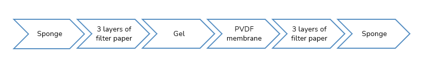
Notice: If detecting small molecule proteins, this step can be omitted, because small molecule proteins can easily diffuse out of the gel. - Cut 6 pieces of membrane and filter paper according to the size of the glue, and put them in the transfer buffer to equilibrate for 10 min. If using PVDF The membrane needs to be saturated with pure methanol for 3-5 s.
-
Assemble and transfer the sandwich:
- Put the sandwich in the transfer box and perform semi-dry or wet transfer according to the instructions of the manufacturer of the blotting equipment.
- After transferring the membrane, cut off the power supply and take out the hybrid membrane.
Immunohybridization and Color Development-Protein Detection:
- Wash the membrane with 25 mL TBS for 5 min, at room temperature, and shake.
- Place the membrane in 25 mL blocking buffer for 1 h, at room temperature, and shake.
- Wash 3 times with 15 mL TBS/T (5 min/T).
- Add primary antibody of appropriate dilution, incubate for 1-2 h at room temperature or overnight at 4°C, shaking slowly.
- Wash 3 times with 15 mL TBS/T (5 min/T).
- Add the appropriate dilution of alkaline phosphatase (AP) or horseradish peroxidase (HRP) labeled secondary antibody, incubate at room temperature for 1 h, shaking slowly.
- Wash 3 times with 15 mL TBS/T (5 min/T).
- Wash once with 15 mL TBS.
- Protein detection (colorimetric method or luminescence method, follow the instructions of the corresponding reagents).
Questions and Answers of Western Blot
Q: What kind of experiment is Western Blotting used for?
A: Western blotting is a conventional technique for protein analysis. It can detect the target protein from the protein mixture, to determine the expression of the target protein in cells or tissues under normal or experimental conditions semi-quantitatively or qualitatively (not quantitative) condition. In addition, Western blotting can be used for subsequent analysis of protein-protein, protein-DNA, and protein-RNA interactions, combined with component separation technology to detect the location of target proteins and become a supplement to immunofluorescence experiments. At the same time, in the post-genomic era, with the continuous improvement of mass spectrometry and protein chip technologies, as a convenient and reliable research tool, Western blotting will cooperate with other technologies to play a more important role.
Q: How to optimize the experimental results of Western Blotting?
A: Western Blotting has many experimental steps, and each step of the operation will have an impact on the final result. During sample preparation, the amount of sample is very important. If the absolute amount of the target protein is small, it needs to be enriched by component separation, IP, and other methods; the amount of sample should be moderate, the right amount will increase the resolution. During electrophoresis, the appropriate gel concentration and voltage and current parameters should be selected to ensure that the proteins are effectively separated and the bands are clear and tidy. When transferring membranes, the appropriate transfer system and current parameters and time should be selected according to the size of the protein. By adjusting the buffer composition, the transfer efficiency and the binding force of the protein and the membrane should be improved. When blocking, you should select the appropriate blocking material and buffer, as well as the concentration of the blocking solution and the blocking time, according to the actual situation.
Reduce non-specific reactions and achieve the highest signal-to-noise ratio. When selecting antibodies, you can try multiple antibodies (monoclonal antibody, polyclonal antibody, or even a combination of multiple antibodies), different dilution ratios, reaction temperature, and time, if possible, to determine the best combination. When developing color, you can try different sensitivities of color developing fluids and signal acquisition systems, and strive to obtain the best results in resolution, contrast, and discrimination.