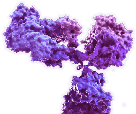
Mouse Anti-Rhesus CD68 Antibody (MO-AB-01564W)
Cat: MO-AB-01564W
Certificate of Analysis Lookup
To download a Certificate of Analysis, please enter a lot number in the search box below. Note: Certificate of Analysis not available for kit components.
Lot Number
To download a Certificate of Analysis, please enter a lot number in the search box below. Note: Certificate of Analysis not available for kit components.
Lot Number
| Size: | |
| Conjugate: | |
| Inquiry |
- Product Details
- Relate Reference Data
Specifications
| Host species | Mouse (Mus musculus) |
| Species Reactivity | Rhesus (Macaca mulatta) |
| Clone | MO01564W |
| Specificity | This antibody binds to Rhesus CD68. |
| Format | Liquid or Lyophilized |
| Storage | Store at 4°C: short-term (1-2weeks) Store at -20°C: long-term and future use |
| Purity | > 90% was determined by SDS-PAGE |
| Purification | Purified with Protein A or G affinity chromatography |
| Cellular Localization | Other locations; Lysosome |
Application Information
| Application | WB, ELISA |
| Application Notes | ELISA: 1:1000-1:3000 Other applications are to be developed. The optimal dilution should be determined by the end user. |
Target
| Introduction | CD68 (CD68 Molecule) is a Protein Coding gene. Diseases associated with CD68 include Granular Cell Tumor and Breast Granular Cell Tumor. Among its related pathways are Hematopoietic Stem Cell Differentiation Pathways and Lineage-specific Markers and Innate Immune System. An important paralog of this gene is LAMP1. |
| Product Overview | Mouse Anti-Rhesus CD68 Antibody is a mouse antibody against CD68. It can be used for CD68 detection in Western Blot, Enzyme-Linked Immunosorbent Assay. |
| Alternative Names | Macrosialin isoform A; CD68 |
| UniProt ID | H9ZFR5 |
| Protein Refseq | The length of the protein is 324 amino acids long. The sequence is show below: MRLAVLFLGALLGLLAAQGTGNDCPHKKSATLLPSFTVTPTATESTGTTSHRTTKSHKTTTHRTTTTGTTSHRPTTATHNPTTTSHRNATVHPTSNSTATSQGPTSSAHPGPPPPSPSPSPASKETIGDYMWTNGSQPCVHLQAQIQIRVMYTTQGGGEAWGISVLNPNKTKVQGSCEGAHPHLLLSFPYGHLSFGFMQDLQQRVVYLSYMAVEYNVSFPHAAQWTFSAQNASLRDLQAPLGQSFSCSNSSIILSPAVHLDLLSLRLQAAQLPHTGVFGQSFSCPSDRSILLPLIIGLVLLGLLALVLIAFCIVRRRPSAYQAL. |
Reference
| Reference | Makori, N., Tarantal, A. F., Lü, F. X., Rourke, T., Marthas, M. L., McChesney, M. B., ... & Miller, C. J. (2003). Functional and morphological development of lymphoid tissues and immune regulatory and effector function in Rhesus monkeys: Cytokine-secreting cells, immunoglobulin-secreting cells, and CD5+ B-1 cells appear early in fetal development. Clinical and Vaccine Immunology, 10(1), 140-153. |
See other products for " CD68 "
| MO-AB-24479R | Mouse Anti-Pig CD68 Antibody (MO-AB-24479R) |
| MO-AB-09878R | Mouse Anti-Cattle CD68 Antibody (MO-AB-09878R) |
| MO-AB-52581W | Mouse Anti-Marmoset CD68 Antibody (MO-AB-52581W) |
| MO-AB-44005W | Mouse Anti-Horse CD68 Antibody (MO-AB-44005W) |
| MO-AB-22684W | Mouse Anti-Chimpanzee CD68 Antibody (MO-AB-22684W) |

Figure 1 Jejunums, ileums, and colons of fetal rhesus monkeys. At day 65 of gestation, macrophages were few, while p55+ DCs were numerous throughout the gut lamina propria, especially at the tips of villi.

For Research Use Only | Not For Clinical Use.
Online Inquiry

